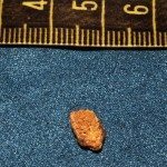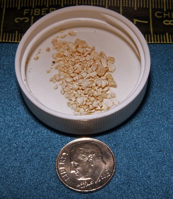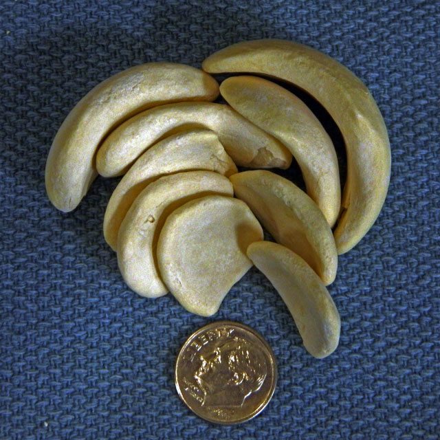
Urinary stone disease (also known as renal stone disease, kidney stone disease, renal calculus disease, Nephrolithiasis, and ureterolithiasis) while found throughout the United States is most commonly found in the “stone belt.” Kidney stones will affect about 1 in 1,000 people. The “stone belt” consists mostly of the southern USA. Kidney stone disease is due to the combination of influences that include diet, heredity, dehydration, and occasionally medication.
Types of Urinary Stones
The most common stones are calcium stones. Calcium stones make up about 70-80% of all kidney stones; however, most people are unaware of the different types of calcium urinary stones that exist. For many years, these were referred to by their mineral or geologic names. These are predominately calcium oxalate dihydrate or weddellite (CaC2O4·2H2O) and calcium oxalate monohydrate or whewellite (CaC2O4·H2O). Others may contain calcium phosphate also known as hydroxyapatite (Ca10(PO4)6(OH)2) or Brushite CaHPO4·2H2O.
Kidney Stones made of Calcium Oxalate Dihydrate

Calcium oxalate dihydrate stones are typically very crystalline in form. Crystalline stones are typically very spiked or rough. If you’ve ever heard anyone refer to their Kidney Stone as a sandspur or cocklebur then they were most likely referring to a calcium oxalate dihydrate stone.
Kidney Stones Made of Calcium Oxalate

Calcium oxalate monohydrate stones on the other hand are usually knobby in shape and have few if any spiked crystals on their surface. The two types of calcium oxalate vary greatly in hardness as well as appearance. The best analogy is that of carbon. While both coal and diamonds are carbon, they exhibit very different chemical and physical properties. Like diamonds, the monohydrate forms are very dense and hard. This hardness must be taken into consideration in choosing a treatment. Calcium dihydrate is much less dense, and the crystalline nature makes them harder to pass but easier to break with Extracorporeal Shock Wave Lithotripsy or ESWL and lasers.
Uric Acid Stone

Uric Acid stone disease is much less common than calcium oxalate stone formation. Uric acid stones make up only 5-10% of all stones. Uric acid causes two different distinct medical conditions. One is stone formation of radiolucent stones. The other condition is gout. Gout is a type of arthritis caused by the crystallization of uric acid crystals in joints. The most commonly affected joints are the big toe and thumb joints. Uric acid stones do not show up on routine radiographs or x-rays such as KUB (Kidney-Ureter-Bladder film) due to their low density. These are characterized to be radiolucent stones. They do show up on CT scan as low density stones in the 400-600 Hounsfield unit range. While they are difficult to localize for ESWL using fluoroscopy, they are very fragile, and when treated, they break very well into multiple small pieces. Pure uric acid stones are usually orange colored stones. Uric acid can also be the nidus or seed crystal that allows calcium stones to form. Once the seed crystal of uric acid forms, calcium is then deposited around this initial uric acid crystal.
Struvite stone disease is most commonly associated with infection within the urinary tract. Struvite stones make up about 10% of all stones. The organisms most likely to cause these stones are urea splitting organisms. As these organisms (germs or bacteria) break down the urea in the urine, the pH of the urine increases from a baseline of pH 5 to a pH of 7, which leads to the precipitation of magnesium ammonium phosphate crystals. The most common bacteria are Proteus mirabilis, Pseudomonas Aeruginosa, Providencia Stuarti, Klebsiella Peumonia, Staphylococcus and Mycoplasma , and Serratia marcescens
Pain From Stones
Passage of a kidney stone or renal calculus is often rated as one of the top 2 pains in humans, which are childbirth and passage of a kidney stone. Women routinely compare passage to labor pains. They often report that labor pains are less intense. The pain is caused by urinary obstruction, not the existence of the stone in the kidney.
Stone disease will affect about 6-9% of all men and until recently 3-4% of women. Recent studies show the gap, between men and women with stones, to be narrowing. The ratio of men to women has changed due to an increased number of stones in women. It has long been noted that the time of first stone formation was after age 18 years. Recently, studies have shown an increase in stones in children as young as 8-10 years old.
Symptoms of Kidney Stones
Symptoms of stone passage include “flank pain”. The flank is the region of your body on your back protected by the last 2 ribs. There may be radiation of the pain around to the lower abdomen on the affected side. Patients frequently experience nausea and occasionally vomiting. As the stone passes out of the kidney into the upper ureter, men may experience testicular pain, and women may have a similar pain in the vagina or groin area. If the stone is very low in the ureter, near the bladder, then there will likely be an onset of frequent urination that can be mistaken for a urinary infection. Bladder infection, cystitis, and UTI are alternate terms for urinary infections. Men may mistake the same symptoms for prostatitis.
Deciding on a Treatment for Kidney Stones
The primary treatment of stones is based on the size and location of the stone. Stones are usually divided, by size, into groups of 1- 4 millimeters (mm), 5-10mm, and stones greater than 10mm. A 6mm stone is about ¼ inch in size. A 12 mm stone is ½ inch in size. There are 25.4 millimeters in one inch. Most stones less than 4 mm will pass and not require surgery. The average time to passage ranges from 3-6 weeks without medical intervention. This can be lessened to as little as 5-10 days with expulsive therapy using alpha blocking medications (Tamsulosin or Flomax, Cardura or Doxazosin, or Hytrin or Terazosin). The alpha blocking medications were first used in medicine as a treatment for hypertension or high blood pressure. These medications lead to dilatation of the ureter and more rapid passage of the stone. Calcium channel blockers such as Procardia (Nifedipine) and steroids such as Prednisone and Methylprednisolone have been used as well.
Surgical Indications for Kidney Stones
Indications for surgical intervention are, having a stone too large to pass, infection behind the stone, intractable pain or vomiting, or complete obstruction of the kidney leading to possible permanent kidney damage. Most of the time, it is acceptable to try to pass the stone if pain and nausea can be controlled and if there is no sign of impending kidney damage.
Risk of Recurrence of Stone Formation
After formation of a stone, there is a 14% risk of having another stone within a year, a 35% risk in the next 2 years, and up to a 52% risk of recurrence at 10 years. This rate of stone recurrence applies if nothing is done to change the patient’s risk. Up to 80-90% of people with a history of stones can modify their risk of recurrent stone formation. One can decrease one’s recurrence rate by change of diet, state of hydration, or by the addition of medications.
Hydration for Prevention
Most people with stones do not drink enough fluids, or what they do drink is high in salt or caffeine. Fluid loss through sweating can also lead to relative dehydration. The goal is to increase your fluid consumption so that there is a 2-liter (2,000-milliliter) or about 2 quarts of output of urine in each 24-hour day. Caffeine is known to be a diuretic and increases urine output. Caffeine also increases the amount of calcium excreted into that urine. This increased calcium excretion leads to more calcium in the urine than can remain in a dissolved form; therefore, crystals begin to form. This initial crystal formation is the beginning of stone formation. The best fluids for stone prevention are lemonade and orange juice. Both juices increase the urinary level of citrate. Citrate has long been known to decrease stone formation. While cranberry juice is widely misused for bladder infections, it can cause stone disease and is not recommended. Water of course is cheap and widely available. In most studies, there appears to be no benefit to bottled water over tap water. It is rare for tap water to contain enough calcium to cause stone formation.
Inheritance of Kidney Stones
Stones commonly occur in families. Most of the time, this history is easily obtainable. Some of your family may have already had a metabolic evaluation, such as a 24-hour urine collection or stone analysis. This information may help other family members as families often make the same type(s) of stones.
Dietary Causes of Kidney Stones
Multiple foods, in excess, have been found to cause stone disease. A partial list is available below.
- Oxalate containing foods.
Oxalate is half of a calcium oxalate stone. Foods increasing urinary oxalate include: chocolate; nuts and nut products; vegetables such as grits, okra, spinach (most dark leafy green vegetables); most berries; draft beer; soy protein; and tea. - Sodium containing foods.
Salt or sodium increases the stone formation. Stone formation risk rises as the salt intake rises. Salt, sodium chloride, should be limited to 2,000mg =2grams of sodium per day. Most Americans consume between 12-15 grams per day. - Calcium containing foods and medications.
Calcium should be eaten in moderation as both very high and very low calcium diets can cause stones. Calcium rich foods include dairy products such as milk, cheese, yogurt, and ice cream. Calcium supplements such as Citracal (calcium citrate), TUMS, and Rolaids (calcium carbonate) increase urine levels of calcium. Spinach is high in calcium and usually increases urine levels of calcium. While more expensive, Citracal (calcium citrate) is the best form of calcium supplement in people with a history of stones needing supplements. Your calcium supplement should include Vitamin D to promote deposition of that calcium in the bones. Citracal, not only supplements your calcium, but the citrate tries to prevent stone formation from any calcium excreted in the urine. While milk substitutes such as soy milk and almond milk have less naturally occurring calcium, they are fortified with calcium and often contain more calcium per serving than cow’s milk contains. Drinking low fat milk does not lower the calcium content. - Protein containing foods.
Protein intake increases the risk of stones. Protein is found in all forms of meat (beef, pork, chicken, and fish) and not just red meat as most patients think. Protein supplements for body builders and the elderly may also lead to stone formation. The Atkins Diet popularized a low carbohydrate, high protein diet. People on this diet noted an increase in stone formation. Protein should be limited to a 4-ounce portion per meal. This portion of meat is about the size of a deck of cards. - Caffeine containing foods and drinks.
Caffeine ingested in any form increases stone formation. This includes coffee, tea, chocolate, energy drinks, caffeine tablets, and soft drinks. Decaffeinated soft drinks contain no caffeine. This is not true of coffee. There is no US government standard for low caffeine. I once read that Starbucks’ decaffeinated coffee still has more caffeine per serving than most other brands of regular coffee contains.
If you have been advised to monitor Oxalate intake with your meals, click here for a 2-page diet plan that lets you know which foods are Little or No Oxalate, Moderate Oxalate, or High Oxalate.
| · Beverages | · Seafood | · Bread / Starch | · Oils |
| · Milk | · Vegetables | · Cakes / Snacks | |
| · Meat / Nuts / Protein | · Fruits | · Ingredients |
Medications Associated With Kidney Stone Formation
The new onset of stones can occasionally be linked to medications. The most common medications are calcium supplements. Studies vary in how much calcium is safe in supplement form. These estimates range from 1,000 to 1,500mg to 2,000mg per day for the prevention of osteoporosis. I usually recommend 1,500mg Calcium citrate with Vitamin D 400-500 IU per day in patients needing supplements.
The commonly used medication Topamax (Topiramate) causes about 1-2% of patients to begin forming stones. Current uses of this medication are for seizure disorders and prevention of migraine headaches. This medication is usually associated with the new onset of metabolic acidosis. This metabolic acidosis in turn lowers urinary citrate levels.
Zonegran (Zonisamide), a medication for control of partial seizures, may also cause 1-2% of patients to begin forming stones. The mechanism is felt to be similar to Topamax by inducing metabolic acidosis.
Vitamin C was popularized by Dr. Linus Pauling, a biochemist not a medical doctor, for the prevention of colds. He recommended 2,000 mg to 5,000mg or more per day. While this has been found not to be useful in preventing colds, it does cause stones at doses above 1,000-2,000mg per day. The excess Vitamin C is converted to oxalate and excreted in the urine. This may lead to stone formation.
Tests For Kidney Stone Recurrence Preventions
- Stone analysis.
Stone analysis breaks down the most common stone’s contents into percentage of the most common minerals in each stone. - 24-Hour urine collection.
A 24-hour urine collection is used to measure the chemicals in the urine to determine which of the above dietary restrictions needs to be applied to you. - Parathyroid hormone measurement.
A parathyroid hormone blood test will test for parathyroid gland over activity. Parathyroid glands are 4 button-sized glands on the surface of the thyroid. These glands regulate calcium in the blood stream and deposition of calcium in bones. If one or more is overactive, the result is breakdown of bone with an increase in urine and blood calcium levels. - Blood chemistry.
Routine serum chemistries are also evaluated to look for illnesses that may be associated with stone formation.
Medications Used for Prevention of Kidney Stones
Urocit-K and Polycitra-K (Potassium citrate) are available in tablet form and can raise urinary levels of citrate enough to decrease the risk of stone formation in many people. While some insurance companies want to substitute cheaper Sodium citrate and Potassium chloride (KCL) for this medication, these are not appropriate substitutes.
Hydrochlorothiazide (HCTZ), a common diuretic used in hypertension, at a dose starting at 25 mg per day, increases urine output while at the same time lowering the calcium content of the urine.
Pyridoxine or Vitamin B6 has been used in the past but with lesser results and has a side effect of neurotoxicity at higher doses.
Magnesium supplementation, Magnesium oxide 400mg per day, may help some patients lower their risk of repeat stone formation.
Allopurinol 300mg per day and Potassium citrate combined with a decrease in protein intake generally makes uric acid stones smaller and less frequent. Uric acid stone disease can usually be more easily controlled than calcium stone disease.
Surgery For Kidney Stones

If medical management fails, then surgery becomes a treatment option. The surgical procedure recommended for you depends on multiple factors including size of the stone, location within the ureter, whether the stone is infected or not, stone density, history of previous surgical results, and history of passage of previous stones.
Open Removal of Kidney Stones
Open surgical removal of ureteral and renal stones, also called ureterolithotomy and nephrolithotomy, is still rarely needed. Starting in the 1980s, newer options greatly reduced the need for this type of surgery.
Extracorporeal Shock Wave Lithotripsy of Kidney Stones
ESWL or Extracorporeal Shock Wave Lithotripsy utilizes a percussion wave generated in water to break the stone. This technology was introduced from Germany in 1985. While the initial procedure submersed the patient in a large tub of water, this machine is rarely used today. Current second and third generation machines push a small self-contained tank of water up against the patient’s side, and the stone is localized in 2 different axes with either x-ray or ultrasound. The same percussion wave technology is then used to fragment or crush the stones into small enough pieces to pass.
Stone Fragments after ESWL

About 80-85% of people will require only one ESWL treatment. If fragments larger than 5 mm remain after lithotripsy, a second treatment may be needed. The shock wave is not an electrical shock but is a percussion wave. Examples of percussion waves most people are familiar with include bomb blasts and depth charges. Think about a bomb blast or a high speed wind blowing out windows or a depth charge cracking the metal in a submarine.
When the lithotripter created percussion wave hits the stone, the stone absorbs the energy and pieces of the stone break off. The more crystalline dihydrate stones are easiest to break. The monohydrate stones are much harder to break. Stone densities can be measured on a CT scan in Hounsfield units. The range of stone density for kidney stones is between 400-1,400HFU. As the density of the stone approaches 1,000HFU, the stone becomes harder to break and may require 2 treatments.
Percutaneous Removal of Renal Stones

Percutaneous stone removal was popularized in the 1980s as a way to avoid incisional or open stone removal. Percutaneous stone removal is used for large stones within the kidney. Usually, these stone are 25 mm or greater in diameter. One inch in maximum diameter equals 25.4 millimeters. The density of the stone also influences the choice of surgery. Denser stones that do not break well with ESWL will respond to mechanical lithotrites (Gyrus, CyberWand, and Microvasive Lithoclast Ultra). These devices use ultrasound to drive a burr or use pneumatic technology to fragment the stone while suctioning out the pieces at the same time.
For this procedure, a small needle is guided through the skin into the kidney through the flank under x-ray guidance. Dilators then enlarge the opening from 2 mm to 10 mm or about 3/8 inches. This avoids the 10-12 inch incision of an open removal. A temporary, plastic sheath is inserted into the newly established access. A telescope is guided down this tract, the stone is broken, and the pieces are removed. A drainage tube or nephrostomy tube may be left in the tract in the flank. This nephrostomy tube is removed after 2-5 days. This procedure is usually used for large stones that might have previously taken multiple ESWL procedures to fragment.
Ureteroscopy for Ureteral Stone Removal
This requires the introduction of a long, thin telescope (both metal and flexible ureteroscopes are available) through the urethra into the bladder and up the ureter to the level of the stone. A quartz, holmium, laser fiber is then typically used to fragment the stone for removal or passage. A ureteral stent, a hollow plastic tube, is then temporarily inserted to prevent swelling that may close the ureter thus causing stone like pain. The ureteral stent is usually removed after 3-5 days depending on each patient’s surgical findings. Unlike metal, vascular stents, ureteral stents must be removed. If left in place for long periods, ureteral stents can encrust with stone crystals and be difficult to remove.
Conclusion
Most people will benefit from a urologic consultation even if they pass their stone. Together, you and your urologist can decide what tests and dietary changes are right for you.